Best Knee Replacement hospital in Delhi: Best Knee Surgeon in India
Life In Motion Clinic is a Pioneer top leading knee replacement hospital in Delhi, India. Dr. Rahul Sharma best knee surgeon having experience of 16+ years at LMC with a team of 16+ Orthopaedic Surgeons. LMC facilitated with the most advanced Orthopaedic facilities. We at LMC aim to treat patients with care without going for Knee surgery. Our Doctor's team aims that knee surgery is the last option. We solve knee-related orthopaedic problems with proper medical treatment and care. So, if facing knee-related pain, or Knee Joint related kinds of health issues, then Book an Online Appointment with Dr. Rahul Sharma best knee surgeon in Delhi, India.
Know the Anatomy of Knee
More About Knee Joint
Do you know the knee is a complex joint? It is made up of or the combination of different structures including ligaments, tendons, bones, and muscles. These different structures work together to maintain the normal functionality of Knee and offer a stable stability that helps the knee to move easily.
Whatever our age 1 month, or 6 month old born or a 50 to 60 years old person, a well functioning and healthy knee responsible for the movement of the whole body. The mobility of the knee helps human beings to participate in various daily activities. The mobility of the knee helps human beings to participate in various daily activities. We at Life In Motion Clinic Orthopaedic Doctors hope that no one can suffer knee-related health problems, and every human being around the world enjoys a healthy life. In life, it happens sometimes human beings stuck with a knee-related problem like Knee pain. Knee pain is the most common problem in old ages, but don't worry, at (LMC) Life In Motion Clinic have a team of knee arthritis doctors near you in Delhi, India. We are just a phone call +91-9811151085 away from you. Dr. Rahul Sharma is available Online to discuss all your Knee related health problems. The knee treatment is necessary, and LMC believes that Knee surgery is not the first and last option. Dr. Rahul Sharma, our head Knee Surgeon, believes that medics, treatments, and physiotherapists solve knee problems without going for knee surgery. So if anyone faces a knee-related health issue, then Book an Online Appointment with the best knee surgeon in Delhi, India.
Know More About Bones in the Knee Joint
You want to know how a knee is made up. It is like a hinge joint made up of 2 bones. These two bones names are the shinbone also known as the tibia and the thigh bone known as the femur.
Do you know at the end of the femur? There are 2 round knobs known as femoral condyles. The femoral condyles articulated with the flat surface of the tibia called or known as the tibial plateau. Well, for the sake of Knowledge, inside of the leg, the tibial plateau known as the medial tibial plateau. on the outside of the leg, The tibial plateau known as the lateral tibial plateau.
Do you know what the patellofemoral groove is? When the 2 femoral condyles form a groove shape structure on the anterior or front side of the knee, then it called the patellofemoral groove.
Do you know what the Kneecap is? The patella is a small bone that sits in this groove known as the kneecap. It is because of this knee cap human being's knee joint protected from direct trauma, so the kneecap act as a shield in our knee joint.
Do you know what the fourth bone called in the Knee joint? Its name is the fibula. The fibula is another bone of the lower leg. This fibula forms a small joint with the tibia. Well, this joint offers a little movement o the knee joint, so it is not considered as the main joint of the Knee.
Articular Cartilage and Menisci
The tibial plateau on the inside of the leg is called the medial tibial plateau, and on the outside of the leg, it is called the lateral tibial plateau.
Movement of the bones causes friction between the articulating surfaces. To reduce this friction, all articulating surfaces involved in the movement are covered with a white, shiny, slippery layer called articular cartilage. The articulating surface of the femoral condyles, tibial plateaus and the back of the patella are covered with this cartilage. The cartilage provides a smooth surface that facilitates easy movement.
To further reduce friction between the articulating surfaces of the bones, the knee joint is lined by a synovial membrane which produces a thick clear fluid called synovial fluid. This fluid lubricates and nourishes the cartilage and bones inside the joint capsule.
Within the knee joint between the femur and tibia, there are two C shaped cartilaginous structures called menisci. Menisci function to provide stability to the knee by spreading the weight of the upper body across the whole surface of the tibial plateau. The menisci help in load bearing by preventing the weight from concentrating onto a small area, which could damage the articular cartilage. The menisci also act as a cushion between the femur and tibia by absorbing the shock produced by activities such as walking, running and jumping.
Ligaments
Ligaments are tough bands of tissue that connect one bone to another bone. The ligaments of the knee function to stabilize the knee joint. There are two important groups of ligaments that hold the bones of the knee joint together, Collateral ligaments and the cruciate ligament.
Collateral ligaments are present on either side of the knee. They function to prevent the knee from moving too far during side to side motion. The collateral ligament on the inside is called the medial collateral ligament (MCL) and the collateral ligament on the outside is called the lateral collateral ligament (LCL).
Cruciate ligaments - This group of ligaments, present inside the knee joint, control the back and forth motion of the knee. The Cruciate ligament in the front of the knee is called anterior cruciate ligament or ACL and the cruciate ligament in the back of the knee is called posterior cruciate ligament or PCL.
Muscles
Muscles: There are two major muscles, the quadriceps and the hamstrings, which enable movement of the knee joint. The quadriceps muscles are in the front of the thigh. When the quadriceps muscles contract, the knee straightens. The hamstrings are in the back of the thigh. When the hamstring muscles contract, the knee bends.
Tendons
Tendons are structures that attach muscles to the bone. The quadriceps muscles of the knee meet just above the patella and attach to it through a tendon called the quadriceps tendon. The patella further attaches to the tibia through a tendon called the patella tendon. The quadriceps muscle, quadriceps tendon and patellar tendon all work together to straighten the knee. Similarly, the hamstring muscles at the back of the leg are attached to the knee joint with the hamstring tendon.
Conditions
-
ACL Tears
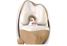 The anterior cruciate ligament, or ACL, is one of the major ligaments of the knee that is in the middle of the knee and runs from the femur (thigh bone) to the tibia (shin bone). It prevents the tibia from sliding out in front of the femur. Together with posterior cruciate ligament (PCL), it provides rotational stability to the knee.
 -
Meniscal Tears
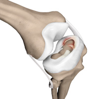 Meniscus tears are the commonest knee injuries in athletes, especially those involved in contact sports. A sudden bend or twist in your knee can the meniscus to tear. This is a traumatic meniscus tear. Elderly people are more prone to degenerative meniscal tears as the cartilage wears out and weakens with age.
 -
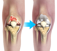
Knee Arthritis
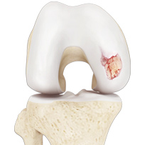 Arthritis is a general term covering numerous conditions where the joint surface or cartilage wears out. The joint surface is covered by a smooth articular surface that allows pain-free movement in the joint. This surface can wear out for several reasons; often the definite cause is not known.
 -
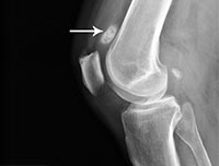
Patellar Dislocation/Patellofemoral Dislocation
 The patella (knee cap) is a protective bone attached to the quadriceps muscles of the thigh by quadriceps tendon. The patella attaches with the femur bone and forms a patellofemoral joint. The patella is protected by a ligament which secures the knee cap from gliding out and is called a medial patellofemoral ligament (MPFL).
 -
Loose Bodies
 Loose bodies are small loose fragments of cartilage or a bone that float around the knee joint. The loose bodies can cause pain, swelling, and locking, and catching of the joint.
Procedures
-
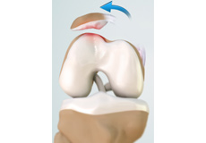
High Tibial Osteotomy
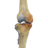 High tibial osteotomy is a surgical procedure performed to relieve pressure on the damaged site of an arthritic knee joint. It is usually performed in arthritic conditions affecting only one side of your knee and the aim is to take pressure off the damaged area and shift it to the other side of your knee with healthy cartilage.
 -
Unicompartmental Knee Replacement
 Unicompartmental knee replacement is a minimally invasive surgery in which only the damaged compartment of the knee is replaced with an implant. It is also called a partial knee replacement.
 -
Total Knee Replacement
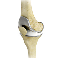 Total knee replacement, also called total knee arthroplasty, is a surgical procedure in which the worn out or damaged surfaces of the knee joint is removed and replaced with artificial parts. The knee is made up of the femur (thigh bone), the tibia (shin bone), and patella (kneecap).
 -
Medial Patellofemoral Ligament Reconstruction
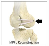 Medial patellofemoral ligament reconstruction is a surgical procedure indicated in patients with severe patellar instability. The medial patellofemoral ligament is a band of tissue that extends from the femoral medial epicondyle to the superior aspect of the patella.
 -
ACL Reconstruction
 The anterior cruciate ligament (ACL) is one of the major stabilizing ligaments in the knee. It is a strong rope like structure located in the centre of the knee running from the femur to the tibia. When this ligament tears, unfortunately, it does not heal and often leads to the feeling of instability in the knee.